Author: Nagla’a A. Abdel-Wahed
Abstract
Introduction:
The presence of an atrophic maxilla creates a serious challenge in cases of implant placement, while maxillary sinus pneumatization further complicates the surgery. This pilot study was performed to investigate the validity of two techniques used to estimate the volumes of bone graft material required in cases that included lateral window sinus augmentation.
Materials and Methods:
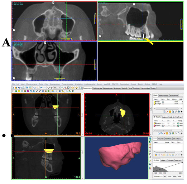
Cone beam computed tomography was used for preoperative volumetric analysis of the maxillary sinus. The analysis was performed using the manual measurement of sinus dimensions, as well as automated measurements via the segmentation technique. The estimated volumes of required bone graft material were compared with actual intraoperative findings in cases requiring lateral window sinus augmentation. For this pilot study, only 5 patients were selected to be included.
Results:
To achieve 80% power and confidence interval of 95%, the sample size should be 35 patients. The correlation coefficient between the segmented volume and mm3 used was – 0.5332, whereas the coefficient between the manual volume and mm3 used was – 0.6784. Consequently, both results indicate that the two methods have a moderate negative correlation with the mm3 used.
Conclusion:
Performing a similar study with an increased number of patients, according to the calculated sample size, increases the possibility of revealing higher correlation between the methods used to analyze the partial volume of the sinus cavity. The estimated sinus volume of the area of augmentation, obtained by using either manual or segmentation techniques, could be considered as a maximum estimate for the required amount of graft material. Furthermore, the segmentation technique may be valuable in preoperative planning of sinus augmentation, as it reveals the topographic shape and morphology of the sinus.